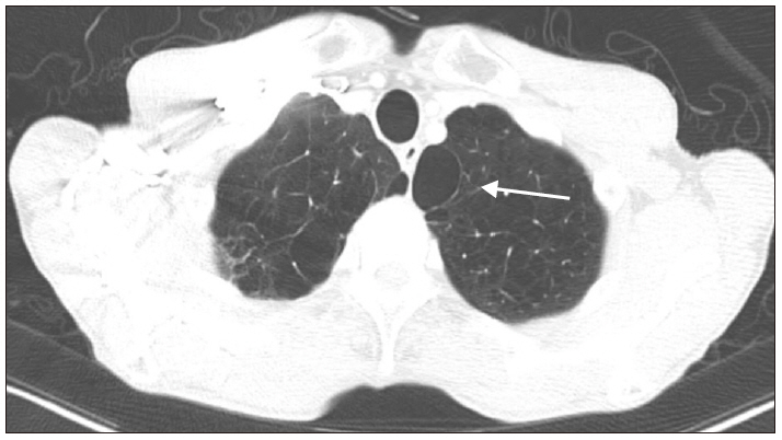
A 75-year-old male weighing 50 kg and having a height of 165 cm was scheduled to undergo right upper lobectomy for the treatment of non-small cell lung cancer. His significant medical history was chronic obstructive pulmonary disease (COPD) and was a 55-pack-year smoker. The preoperative pulmonary function test showed a moderate obstructive pattern with a forced expiratory volume of 1.64 L (61% of predicted) in one second. The chest computerized tomography revealed subsegmental atelectasis in bilateral lower lobes and right middle lobe, emphysema with bullae in bilateral lungs (
Fig. 1), and scanty right pleural effusion. An electrocardiogram (ECG) performed before the surgery showed right bundle branch block, and transthoracic echocardiography revealed normal left ventricle (LV) global systolic function (ejection fraction [EF] = 58%) and abnormal relaxation of LV filling pattern.
The patient received 0.2 mg of glycopyrrolate intramuscularly as a premedication. On arrival in the operating room, intraoperative monitoring was performed including ECG, non-invasive blood pressure, pulse oximetry, bispectral index, and capnography. The immediate preoperative vital signs of the patient were as follows: systolic/diastolic blood pressure of 112/74 mmHg, a heart rate of 90 beats/min, and Oxygen saturation (SpO2) of 96%. After preoxygenation with 100% oxygen, general anesthesia was induced with intravenous 70 mg of propofol, 20 mg of lidocaine, and continuous infusion of remifentanil. After confirming adequate mask ventilation, 60 mg of succinylcholine was given to facilitate passage of a double-lumen endotracheal tube (DLT), after which neuromuscular blockade was maintained with intermittent boluses of intravenous rocuronium. The trachea was intubated with a 37 Fr. left-side DLT (Mallinckrodt™, Covidien Inc., Ireland) under direct laryngoscopy. The DLT position was confirmed by chest auscultation and fiberoptic bronchoscopy. A left radial artery catheter and a central venous catheter were placed via the right internal jugular vein. Arterial blood gas analysis (ABGA) during two-lung ventilation (FiO2 0.6/end-tidal CO2 concentration [EtCO2], 34 mmHg) revealed pH 7.369, PaCO2 40.6 mmHg, PaO2 256.3 mmHg, and SaO2 99.8%. The patient was placed in the left lateral decubitus position for the planned video-assisted thoracoscopic surgery (VATS), and DLT positioning was again confirmed with fiberoptic bronchoscopy.
General anesthesia was maintained with balanced anesthesia using oxygen-air-sevoflurane (1-2 vol%) and continuous infusion of remifentanil. After endotracheal intubation, 40 mg of rocuronium was administered and an additional dose of 10 mg was administered twice until the event described below occurred. Mechanical ventilation during OLV was initiated using pressure-controlled ventilation-volume guaranteed mode with a tidal volume of 300 ml and a respiratory rate of 14 breaths/min. After about 30 min of procedure, VATS was converted to thoracotomy due to surgical site adhesion. Peak airway pressure (PAP) was 14 cmH
2O during two-lung ventilation. After OLV, the PAP was 25 cmH
2O initially and gradually increased to 35 cmH
2O within 10 min. SpO
2 gradually decreased from 97% to 80% during the next 5 min. As a rescue measure, two-lung ventilation was performed for several minutes and SpO
2 increased to 96%. After reapplication of the OLV, SpO
2 gradually decreased from 98% to 70%. Inspired oxygen was increased from 60% to 100%, and two lung ventilation was reinstituted. Oxygen saturation increased to 95% for approximately 5 min. Systolic blood pressure decreased to 70 mmHg; subsequently, 100 µg of phenylephrine was administered twice and infused continuously. However, blood pressure was maintained as 85/60 mmHg. The ABGA (FiO
2 1.0, EtCO
2 23 mmHg) was checked and the outcomes were pH 7.223, PaCO
2 60.9 mmHg, PaO
2 61.9 mmHg, and SaO
2 85.6%. Dopamine infusion (10 µg/kg/min) was started and systolic blood pressure was maintained above 100 mmHg. With intermittent suction of the DLT, oxygen insufflation into the non-dependent lung and dopamine administration, SpO
2 was maintained between 85-90% during OLV. Subsequently, the PAP was noted to be about 35 cmH
2O (maximum 37 cmH
2O). We did not apply positive end-expiratory pressure to the dependent lung because of the high PAP. Nearly 2 h after the operation, systolic blood pressure suddenly dropped below 80 mmHg and fell below 50 mmHg within 1 min, SpO
2 decreased from 90% to 70%, and EtCO
2 decreased to 17-18 mmHg. Almost immediately, the arterial waveform disappeared and asystole was revealed on ECG. Two-lung ventilation was reinstituted and the surgical site was temporarily closed using dressing films. The patient was placed in the supine position and resuscitated with atropine and epinephrine, and the surgeon started closed cardiac compression. At the same time, we asked for help in applying ECMO. About 17 min later, recovery of spontaneous circulation achieved. Nearly at the same time, veno-arterial (VA) ECMO was also started via the right common femoral artery and common femoral vein. On auscultation, lung sound of the left lung field was decreased markedly. Tension pneumothorax of the dependent left lung was suspected. Portable chest X-ray revealed tension pneumothorax of the left lung (
Fig. 2) and a chest tube was inserted immediately. Follow-up chest X-ray showed re-expansion of both the lungs (
Fig. 3). The patient was returned to the left lateral decubitus position, and the right upper lobectomy was performed after the right lung was selectively collapsed. At this time, the left lung was ventilated with a tidal volume of 250 ml and a respiratory rate of 13 breaths/min, and PAP was about 35 cmH
2O. After completion of the surgical procedure, the DLT was replaced with a single lumen endotracheal tube of 7.5 mm internal diameter, and the patient was hemodynamically stable. He was transferred to the intensive care unit while maintaining VA-ECMO.
Immediate postoperative echocardiography revealed moderate LV global and severe right ventricle (RV) free wall motion abnormality and decreased LV systolic function (EF = 40%). On the 10th day, transthoracic echocardiography showed normal LV global wall motion but still severe RV free wall motion abnormality was apparant. On the 14th day, echocardiography revealed normal RV free wall motion. Finally, ECMO was weaned off and the patient’s trachea was extubated.


