CASE REPORT
A 3-year-old boy (height 103 cm, weight 24 kg) from Dubai with known G6PD deficiency (class IV or V) visited the emergency room with complaints of abdominal pain lasting one week. The pain varied in intensity, occurred primarily at bedtime, and was associated with vomiting and nausea. A computed tomographic scan of the abdomen revealed right hydronephrosis with a severely dilated renal pelvis, suggesting complete right ureteropelvic junction obstruction. Surgical intervention via right pyeloplasty was planned to eliminate the obstruction and preserve the remaining function of his right kidney. There was no history of hemolysis, jaundice, or blood transfusion. All routine preoperative parameters were normal. His hemoglobin (Hb) level was 12.7 g/dl; however, other hemolytic laboratory parameters were not evaluated because there was no history of G6PD deficiency-related sequelae such ashemolysis, jaundice, or blood transfusion, and clinical examination revealed no abdominal tenderness. Intraoperative monitoring included electrocardiogram, oxygen saturation (SpO
2), non-invasive blood pressure, arterial blood pressure, end tidal carbon dioxide (ETCO
2), and temperature. The patient underwent 3 min of denitrogenation with 100% oxygen through a facemask. Anesthesia was induced using thiopental sodium (120 mg) with sevoflurane in oxygen. After confirming loss of consciousness, rocuronium (12 mg) and fentanyl (25 μg) were administered. Appropriate mask ventilation with ETCO
2 monitoring was initiated to avoid hypercapnia. Endotracheal intubation was then performed using a 4.5-mm tube (internal diameter) with an inflatable cuff. After applying the ventilator, ETCO
2 was 29 mmHg and peak inspiratory pressure (PIP) was 19 cmH
2O. The initial tidal volume was 240 ml and respiratory rate was 15 breaths/min. Anesthesia was maintained with sevoflurane in oxygen. The patient was placed in the left lateral decubitus position for surgery. Cefotaxime 500 mg was administered as a prophylactic antibiotic. Before an incision was made, fentanyl (25 μg) was given for analgesia and rocuronium (2.5 mg) for abdominal relaxation. We also applied a heated circuit and air warmer to keep the patient warm. After the surgeons placed some ports, the abdomen was insufflated with CO
2 at a pressure of 10 mmHg. The PIP and ETCO
2 were monitored carefully during the CO
2 insufflation period and did not increase much. We performed a blood gas analysis each time the ventilator setting was changed (
Table 1).
Table 1
Respiratory Parameters and Results of Arterial Blood Gas Analysis during Surgery
*
|
After induction |
During CO2 insufflation |
Outside CO2 insufflation |
|
CO2 (mmHg) |
0 |
10 |
0 |
|
TV (ml) |
240 |
240 |
240 |
|
RR (breaths/min) |
15 |
16 |
15 |
|
FIO2
|
1.0 |
0.5 |
0.5 |
|
PIP (mmHg) |
19 |
22-26 |
15 |
|
ETCO2 (mmHg) |
29 |
30-32 |
28-29 |
|
pH |
7.48 |
7.43 |
7.41 |
|
pCO2 (mmHg) |
31.0 |
33.0 |
33.0 |
|
pO2 (mmHg) |
437 |
283 |
282 |
|
Base excess (mEq/L) |
0.1 |
−1.8 |
−3.0 |
|
Bicarbonate (mEq/L) |
23 |
22 |
21 |
|
SpO2 (%) |
100 |
100 |
100 |
|
Glucose (POCT, mmol/L) |
5.61 |
8.38 |
10.71 |
The duration of anesthesia was approximately 4 h and its course was uneventful. Residual neuromuscular blockade was reversed using glycopyrrolate (0.2 mg) and pyridostigmine (2 mg), and the patient was extubated. The day after surgery, laboratory investigations revealed an Hb level of 12.0 g/dl; bilirubin, 0.4 mg/dl; haptoglobin, 116 mg/dl; reticulocytes, 1.5%. Additional tests of hemolysis parameters, such as lactate dehydrogenase, unconjugated bilirubin, and peripheral blood smear were not performed because there were no primary signs of hemolysis. The color and volume of the perioperative urine were normal. Postoperative pain was controlled using pethidine. The patient was discharged two days after surgery, owing to his improved condition.
DISCUSSION
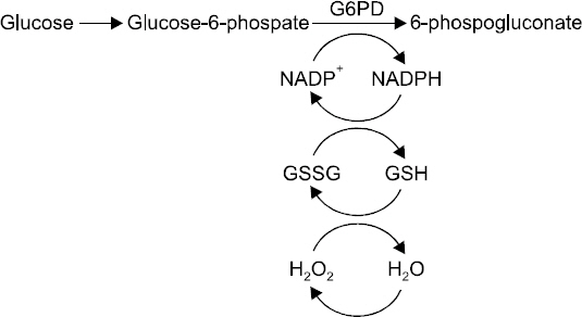
G6PD is an enzyme that catalyzes the first reaction in the pentose phosphate pathway (PPP). The PPP reduces nicotinamide adenine dinucleotide phosphate (NADPH), which enables the preservation of the reduced form of glutathione. Reduced glutathione acts as a scavenger for oxidative metabolites in cells (
Fig. 1). Because RBCs do not contain mitochondria, the PPP provides the only source of NADPH; therefore, RBCs are especially susceptible to oxidative stress, resulting in hemolytic anemia [
8]. Hemolysis can occur after exposure to drugs or other substances that produce peroxides and other oxidizing radicals. When hemoglobin is oxidized, it loses much of its function.
Fig. 1
Pentose phosphate pathway. G6PD: glucose-6-phosphate dehydrogenase, NADP+: nicotinamide adenine dinucleotide phosphate, NADPH: reduced NADP, GSSG: oxidized glutathione, GSH: reduced glutathione, H2O2: hydrogen peroxide.

During surgery, anesthetic management should focus on minimizing oxidative stress, and monitoring and treating hemolysis. The main causes of oxidative stress are the consumption of fava beans, certain drugs, infections, and metabolic conditions such as diabetic ketoacidosis [
8,
9].
The monitoring of temperature and blood gas analysis, in addition to the usual parameters (electrocardiography, non- invasive blood pressure, SpO
2), are important for the prevention of hypothermia and the detection of acidosis and hyperglycemia [
2]. In this case, we inserted an esophageal stethoscope and arterial catheter, and monitored excreted urine to detect hemoglobinuria as a sign of active hemolysis. Our patient underwent robot-assisted laparoscopic pyeloplasty (RALP), which involved prolonged intraperitoneal CO
2 insufflation. When CO
2 insufflation is initiated, PIP increases and CO
2 absorption leading to acidosis occurs [
6,
7], which in turn is a potential precipitating factor in hemolysis [
2]. Moreover, the patient’s body mass index was 23.18 kg/m
2 (103 cm, 24 kg), which contributed to an elevation in PIP. Although laparoscopic surgery is not suitable for G6PD-deficient patients, RALP overcomes some of the disadvantages of traditional laparoscopy, including long operative times [
10]. Robotic surgery provides three-dimensional imaging, thereby allowing better visualization [
11]. Therefore, the CO
2 insufflation pressure can be minimized. Usually, the abdomen is insufflated with CO
2 at a pressure of 10-15 mmHg in RALP. To avoid adverse events, the surgeons inflated the abdomen with CO
2 at a pressure of 10 mmHg. In addition, we attempted to intubate with a larger size tube and minimized the head-down position to prevent the PIP from increasing. After the child was placed in the left lateral decubitus position, the PIP was not elevated significantly. During the operation, ETCO
2 was maintained at 29-32 mmHg, and PIP was maintained at 22-26 cmH
2O during CO
2 insufflation, and at 15-19 cmH
2O otherwise. Minute ventilation was adjusted in accordance with the fluctuations in CO
2 levels, to maintain the pH, base excess, bicarbonate, and partial pressure of CO
2 within normal ranges. We performed blood gas analyses three times over a 4 h period (
Table 1); all parameters were in the normal range. Typically, laparoscopic surgery makes the patient’s condition more acidic [
6,
7]. In this case, we adopted various measures in an effort to prevent this from occurring, including minimizing CO
2 insufflation pressure, using a larger-sized tube, maintaining appropriate ventilation, and monitoring arterial blood gas composition.
Drugs that cause oxidative stress and/or induce methemoglobinemia should be avoided in G6PD-deficient patients [
4]. Seven currently used medications, including dapsone, methylthioninium chloride (methylene blue), nitrofurantoin, phenazopyridine, primaquine, rasburicase, and tolonium chloride (toluidine blue), have been contraindicated with convincing supportive evidence [
4]. Methylene blue is commonly used in urosurgery to ensure ureteral patency. However, when the surgeon in this case requested intravenous methylene blue during the procedure, we advised against it. The NADPH MetHb reductase system continuously converts methemoglobin (MetHb) to hemoglobin in RBCs. Methylene blue acts as a cofactor for NADPH MetHb reductase and is therefore used for the treatment of methemoglobinemia. Because of low levels of NADPH, G6PD- deficient patients are at increased risk for methemoglobinemia; hence, methylene blue is ineffective. In general, there is insufficient evidence regarding medication use, including that of anesthetic agents, in G6PD-deficient patients. One in vitro study revealed that isoflurane, sevoflurane, diazepam, and midazolam have an inhibitory effect on G6PD activity [
5]. However, sevoflurane and midazolam are controversially discussed in one review, and there are some case reports of the safe use of sevoflurane in G6PD-deficient patients [
3,
4]. Our experience in this case suggests that sevoflurane can be considered as safe. The urine color was normal, and the patient’s temperature was maintained at 36.8-37.4°C. It is usually difficult to detect hemolysis in patients under general anesthesia. Free hemoglobin in the urine (dark in color) can be a clue [
12]. During surgery in this particular case, however, signs of hemolysis were not detected.
The risk of infection is another important consideration [
13]. Although the contributing factors are not known, infection is a common risk factor for hemolysis in G6PD-deficient patients. In this case, cefotaxime (500 mg) was administered as a prophylactic antibiotic.
In general, hemolysis becomes evident after 1 to 3 days in patients with risk factors, and anemia worsens until approximately day 7. Although acute hemolysis is self-limiting, in rare cases it can be sufficiently severe to warrant blood transfusion [
12]. The symptoms of hemolysis include cyanosis, headache, fatigue, tachycardia, dyspnea, lethargy, lumbar/substernal pain, abdominal pain, splenomegaly, hemoglobinuria, and/or scleral icterus [
14]. Peripheral blood smear microscopy may include RBC fragments such as schistocytes and reticulocytes, and Heinz bodies. Blood levels of lactate dehydrogenase and unconjugated bilirubin will be elevated, and haptoglobin levels decreased. Hemosiderin and urobilinogen can be present in the urine. To rule out an immunological reaction, a direct Coombs test should yield negative findings [
12].
In summary, the inadequate management of G6PD-deficient patients increases the risk of them developing acute hemolytic anemia, which can lead to permanent neurological damage or death. Avoiding oxidative stress and detecting and monitoring hemolysis are key strategies for the successful anesthetic management of such patients. Although patients undergoing robot-assisted laparoscopic surgery are susceptible to acidosis and hemolytic conditions, the patient in this case experienced no adverse events with adequate monitoring and management. Additionally, there is no evidence-based consensus regarding the use of anesthetic agents in patients with G6PD deficiency. In our case, thiopental sodium, sevoflurane, fentanyl, pethidine, and rocuronium were found to be safe.


